Why X-rays are taken at all
Dental X-rays play an important role in any treatment plan. A range of examinations — preventive and diagnostic — give clinicians a better understanding of each patient's anatomy and needs. The companion article How safe are dental X-rays? covers the same ground from the dose side; this one is about the imaging types and where the safety threshold actually sits.
Imaging helps diagnose gum disease, tooth and gum infections, cavities, and assess bone density.
Without imaging, some conditions would be left undiagnosed and therefore untreated.
That is the justification for the dose, and it is a real one: decay between teeth and bone loss below the gum are not visible on examination. A dentist working without imaging is diagnosing with a significant part of the picture missing. An abscess at a root tip is the clearest example — it can be entirely silent and entirely invisible, and can affect your general health if it is left.
The two categories
Intraoral — captured inside the mouth
High detail at specific sites — cavities, tooth roots, developing teeth.
- Bitewing X-rays — to check between teeth, and assess restorations. This is the routine image taken at a check-up, and what finds the decay that could still be reversed
- Periapical X-rays — close-up views of individual teeth including their roots. These are what root canal treatment is planned and checked against — see everything you need to know about root canal treatment and why the microscope matters
- Occlusal — to assess the bite relationship between upper and lower teeth; see what is malocclusion of the teeth?
Extraoral — captured outside the mouth
A larger area in one exposure, covering all the teeth, jaw and oral anatomy.
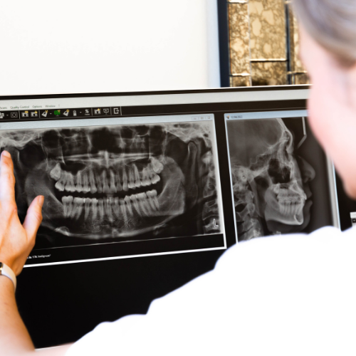
- Orthopantomogram (OPG) — a full mouth X-ray. This is what shows wisdom teeth position and unerupted teeth
- Cephalometric imaging — X-rays of the head and facial profile, used in orthodontic planning and before jaw surgery; see what is orthognathic surgery?
- Cone Beam Computed Tomography (CBCT) — 3D imaging of specific areas of the face and teeth. Its main use is implant planning, where bone volume and nerve position have to be known before surgery — see what do I need to know about dental implants? and bone grafting
CBCT carries a higher dose than a plain film, which is why it is reserved for cases where the three-dimensional information changes the plan. The equipment in the building is listed under technology.
What each one costs you in dose
The categories above are not interchangeable, and neither are their doses. The International Atomic Energy Agency publishes typical effective doses by examination type, which is the most useful way to read the rest of this page:
- Intraoral dental X-ray: 1–8 μSv
- Panoramic examinations: 4–30 μSv
- Cephalometric examinations: 2–3 μSv
- CBCT: 50 μSv or below for small or medium scanning volumes, and 100 μSv for large volumes (based on median values from the literature)
One microsievert (μSv) is a thousandth of a millisievert (mSv), so the whole list sits between 0.001 and 0.1 mSv. The practical point is the spread: a large-volume CBCT can carry something in the order of a hundred times the dose of a small intraoral film. The IAEA also notes that rapid technological improvements to CBCT equipment mean typical dose ranges are likely to change, so an older published figure for CBCT is more likely to overstate than understate.
How exposure is controlled
Great care is taken to ensure X-rays are taken only when necessary, and kept to a minimum — which keeps the total dose low if you need several during treatment. That matters most in long, staged treatment — see complex dental cases: what happens when multiple specialists collaborate.
Your dentist selects the most appropriate imaging for your needs, and explains why — see understanding your treatment.
If an extraoral radiograph is required, you may be referred to an imaging clinic, where a radiographer performs the examination using the lowest dose that still produces a high-quality image.
Radiation safety is taken seriously at every step — exposure selection, collimation (the precision of the beam), and lead shielding.
Collimation is the one worth knowing about. It narrows the beam to the area being imaged, so the surrounding tissue is not exposed at all.
The numbers, in context
Background radiation
We are exposed to radiation every day in the natural environment. The amount varies with soil, rock, altitude, latitude and diet.
In Australia the average is approximately 1.5 mSv per year from natural sources — roughly equivalent to 75 chest X-rays.
That comparison is the useful anchor. Simply living in Australia delivers the equivalent of 75 chest X-rays annually, before any medical imaging at all. Both of those figures are Smile Solutions' own; they are not drawn from the IAEA or ARPANSA material cited elsewhere on this page, and if the exact numbers matter to your decision, ARPANSA is the body to check them against.
A dental X-ray
A single dental exposure delivers between 0.001 and 0.005 mSv.
That is comparable to:
- The radiation received on a one- to two-hour flight
- About one day of natural background exposure
The IAEA makes the same comparison independently, and adds the detail that the examination type changes the answer: doses from intraoral and cephalometric procedures are usually less than one day of natural background radiation, while panoramic doses are more variable, and even at the high end of the range are equivalent to a few days of natural background radiation — similar to a chest radiograph. So the figure above is an intraoral one; a panoramic image belongs further up the scale, and a CBCT further up again.
Conventional 2D dental X-rays emit very small doses and are considered very safe. Digital X-rays use very low doses — dental imaging uses the lowest dose of all plain-film radiography. What digital sensors changed, and why older published figures overstate the dose, is covered in the benefits of holistic dentistry.
Where harm begins
Radiation becomes harmful at very high doses — above around 500 mSv — and reactions occur shortly after exposure.
Those doses arise only from large nuclear or radiation accidents. They do not arise from plain film radiography.
To put the gap in perspective: 500 mSv is roughly 100,000 to 500,000 times a single dental exposure.
The published literature describes the same threshold in technical terms. Radiation effects are divided into two kinds: deterministic effects, which require a high dose and result from killing cells, and stochastic effects, which result from damaging DNA. On the first of those, a 2023 review of dental radiation exposure indexed in PubMed Central states plainly that conventional dental imaging never causes deterministic effects. That is the sourced version of the paragraph above, and it is a statement about the dose threshold — not about the second category.
The honest qualification
None of this means the dose is zero. There is a recognised relationship between radiation dose and cancer risk, which is why the principle of keeping exposure as low as reasonably achievable governs all imaging. That is the stochastic side, and it is the one that does not have a threshold below which it disappears.
What it means is that the dose from a dental X-ray is very small, and the decision to take one is a judgement that the diagnostic information is worth that small exposure.
The questions worth asking are therefore about necessity rather than safety:
- Why is this image needed?
- Which examination is it, and what is the dose for that examination? — the IAEA ranges above vary by a factor of about a hundred across the list
- How often do I need them? — the interval should reflect your decay and gum risk, not a fixed schedule. The same logic applies to how often you should see a dentist at all
- Are recent images available from another practice? Continuity makes this easier — see is it important to have a family dentist? — and it applies equally when you are seeking a second opinion
And tell your dentist if you are pregnant or may be, so that decision is made knowingly — see is it safe to visit the dentist during pregnancy? and oral health care while pregnant.
For children the same indication-based rule applies, and it is one of the things a paediatric dentist is trained to judge — see children's dentistry.
If you have any concerns about your imaging, your dentist or radiographer will discuss them with you beforehand. More questions of this kind are collected in 5 questions you've always wanted to ask your dentist.
Common questions
Is the calculation different for a child?
Yes, in both directions, and the guidance has changed in a way most parents have not been told about.
Children are more radiosensitive than adults. A 2023 study of 4,455 intraoral radiographs taken in children and adolescents at a German university hospital, indexed in PubMed Central, sets out why: "the higher cell division rate, the higher proportion of water in children's tissues, the close proximity of radiation-sensitive organs (e.g., lens, pituitary and thyroid glands and oral cavity) and the longer expected lifetime after exposure."
But the doses involved are also small. In that dataset the average effective dose was 0.77 μSv for intraoral dental and bitewing radiographs and 2.22 μSv for occlusal radiographs — at or below the low end of the IAEA adult range quoted above.
The rule that changed is about routine imaging. X-ray screening on a schedule is no longer recommended: the European Association of Paediatric Dentistry now calls for "an individualized and patient-specific justification for X-ray diagnostics as best clinical practice", and the American Academy of Pediatric Dentistry states that "the timing of the radiographic examination should not be based upon the patient's age, but upon each child's individual circumstances." The governing principle has been restated in the same spirit — from ALARA (as low as reasonably achievable) to ALADA (as low as diagnostically acceptable) and then ALADAIP, which adds "being indication-oriented and patient-specific".
And the paper does not pretend the dose is nothing. It reports a biomarker study in which "bitewing radiographs produced a threefold increase in micronuclei, while digital panoramic radiographs produced a twofold increase" in cells taken from the cheek lining of children days afterwards — micronuclei being a marker of genetic damage, not a disease. The authors' own conclusion is the one to take: "with the understanding that intraoral dental radiographs are not harmless, it is of particular importance to establish diagnostic reference levels for use in pediatric dentistry." There are, as yet, no such reference levels for children.
The practical version: ask what this particular image is for, and expect an answer about your child rather than about their age.
How much does the image actually change the diagnosis?
More than nothing, less than everything — and the literature is genuinely split, which is worth knowing before you agree or decline.
On the side of imaging: the same 2023 paper reports that "visual clinical examination accompanied by intraoral dental or bitewing radiographs detected up to 50% more proximal caries lesions in the posterior primary and permanent teeth compared to visual examination alone", and that without radiographs "the caries prevalence would be underestimated".
On the other side: the paper also records that Pontes and colleagues "questioned the benefit of radiographs as a protocol in the diagnostic process to detect caries in children", observing "only modest changes in the treatment decisions regarding primary molars made after additional radiographic evaluation compared to exclusively visual examination".
Both findings can be true, because detecting more lesions and changing more decisions are different things. The standard that reconciles them is the one the paper states: a justified radiograph "should make a substantial contribution to distinguishing between treatment options". If the image will not change what happens next, it is not justified.
There are also radiation-free options for some of this work. The paper names near-infrared transillumination, laser fluorescence, fibre-optical transillumination and other fluorescence-based cameras as alternatives that "might be considered to enhance the accuracy of visual caries diagnostics". They do not replace a radiograph for everything — bone level and root tips are still radiographic questions — but they are a reasonable thing to ask about.
Can I decline an X-ray?
Yes, and the literature is clear that you are being asked, not told. The 2023 review states the obligation on the practitioner: "dentists have to justify every radiograph for every patient individually, weighting the potential benefit (information to change treatments) against the potential radiation risk."
What that gives you is a right to the reasoning rather than a right to a better answer. If the reason offered is "we do them every two years", that is a schedule, not a justification, and it is fair to ask what this one is expected to show.
It cuts the other way too, and the same paper says so in a single sentence worth quoting in full: "there is a certain health risk associated with every X-ray. It is our duty to keep this risk as low as possible. However, no X-ray should be withheld from the patient that is necessary for appropriate medical or dental care."
Declining an image is a decision with consequences — usually that a diagnosis is made with less information, or deferred. Ask what would be missed, and what the plan is if you say no.
What is actually being exposed, and does the thyroid collar do anything?
The tissues near the beam, at very small absorbed doses. For an intraoral dental radiograph, the 2023 review summarises published organ doses as roughly 0.63 μGy to bone marrow, 2.36 μGy to the brain, 7.97 μGy to the thyroid gland and 22.79 μGy to the salivary glands. Larger examinations scale up: an occlusal view of the upper jaw has been measured at around 70 μGy to the brain and 50 μGy to the salivary glands, and a full-mouth bitewing examination at more than 100 μGy in the salivary glands and more than 150 μGy in the oral mucosa.
Note which organ tops that list. It is the salivary glands, not the thyroid — the beam passes through the floor of the mouth and the cheeks.
On shielding and technique, the paper lists what actually reduces exposure for intraoral radiography: "rectangular collimation, fast image receptor speeds and thyroid shielding". Rectangular collimation is the one that does the heavy lifting, because it shrinks the irradiated area rather than blocking a beam after the fact. It is a fair question to ask whether the machine being used is rectangularly collimated.
Is a digital X-ray genuinely lower dose than the old film?
Yes, and this is one of the few places where the improvement is described in the literature without hedging. The 2023 review states that "the switch from film-based to digital X-ray machines led to a drastic improvement, lowering the effective dose and, consequently, the absorbed organ doses".
The practical consequence is that published dose figures age badly in one direction only. A number quoted from a film-era paper overstates what a current digital machine delivers — which is worth remembering when you encounter an alarming figure online without a date attached to it.
Two further levers sit with the operator rather than the technology: the tube voltage and exposure time selected, and the size of the sensor used. In the paediatric dataset above, the machines ran at 60 kV, described as "the lowest value recommended for intraoral radiographs", with an exposure time of 0.1 seconds and small 2.2 × 3.5 cm sensors for children.
Related reading
- How safe are dental X-rays?
- What is holistic dentistry?
- Dental myths exposed
- Oral cancer: how your dentist can help with early detection
- Why would I need to see a dental specialist?
- Finding a dentist online in Australia
- General dentistry and specialist care
Practical details
We have on-site OPG and CBCT imaging — see technology and how to find us.
Smile Solutions, Level 1, 220 Collins Street, Manchester Unity Building, Melbourne VIC 3000. Phone 13 13 96, or theteam@smilesolutions.com.au; full details on the contact page. Monday–Friday 8.00am–6.00pm, Saturday 8.30am–1.30pm, Sunday by appointment. The clinicians are listed on our team.
Published 2 October 2018. Dose figures are approximate and vary with equipment and technique. Figures attributed to the IAEA are typical effective doses for the examination named, not a measurement of any particular machine. Organ-dose and paediatric figures are drawn from published studies of other patient populations and equipment, not from measurements of this practice. General information only; it does not replace advice from your treating practitioner.
Smile Solutions trades under ABN 28 193 514 103.
Images on This Page
-
https://www.facebook.com/tr?id=800557773428361&ev=PageView&noscript=1
(no alt text)
-
https://www.smilesolutions.com.au/wp-content/uploads/2025/10/calendar_month.svg
(no alt text)
-
https://www.smilesolutions.com.au/wp-content/themes/arcadian-wordpress-theme/assets/images/smile_solutions_logo_white.png
Smile Solutions Logo
-
https://www.smilesolutions.com.au/wp-content/themes/arcadian-wordpress-theme/assets/images/smile_solutions_logo_purple.png
Smile Solutions Logo
-
https://www.smilesolutions.com.au/wp-content/uploads/2023/11/SS-Tooth-1-1-150x150.png
Smile Solutions
-
https://www.smilesolutions.com.au/wp-content/uploads/2025/09/Wisdom-Teeth-Smile-Solutions.png
Smile Solutions Dentist Melbourne
-
https://www.smilesolutions.com.au/wp-content/uploads/2025/09/Root-Canal-2-300x217.jpg
Root Canal
-
https://www.smilesolutions.com.au/wp-content/uploads/2026/08/Mouth-Breathing-300x270.jpg
Mouth Breathing
-
https://www.smilesolutions.com.au/wp-content/uploads/2025/09/Dental-Crown-at-Smile-Solutions-300x300.jpg
Dental Crown at Smile Solutions
-
https://www.smilesolutions.com.au/wp-content/themes/arcadian-wordpress-theme/assets/images/family_owned_bussiness.png
Family Owned Business
-
https://www.smilesolutions.com.au/wp-content/themes/arcadian-wordpress-theme/assets/icons/icon--phone.svg
Smile Solutions Contact
-
https://www.facebook.com/tr? id=800557773428361&ev=PageView&noscript=1
(no alt text)