Why is the microscope so crucial in endodontic treatment?
The gap between reputation and results
Endodontic treatment — root canal treatment — has often been described as the epitome of discomfort, a reputation kept alive by urban legend more than by current practice. See Root Canal.
So it is worth putting the number first:
Studies show endodontic treatment has a success rate of over 90 per cent.
The interesting question is what accounts for the gap between that figure and the treatment's reputation. A large part of the answer is what the operator can actually see.
What the published figures actually say
Success and survival are two different measurements, and most of the confusion about root canal statistics comes from mixing them.
A 2025 review of endodontic outcomes in the British Dental Journal defines a favourable outcome as the absence of symptoms and clinical signs of disease — such as mobility, a sinus tract or a probing defect — with no loss of function, and, on a radiograph, an intact apical periodontal ligament space with any previous periapical radiolucency resolved. Survival asks something simpler: is the tooth still there, and still working? A tooth can therefore survive for years and still be counted as an endodontic failure.
The long-run figures. A private-practice cohort of 598 root-treated teeth in 312 patients, followed for a mean of 21 years and published in the peer-reviewed literature indexed on PMC, reported:
- overall success of 87.8% at tooth level (95% CI 84 to 90%), and 80.8% at patient level (95% CI 75 to 86%)
- cumulative success of 93% at 10 years, 85% at 20 years and 81% at 30 years
- cumulative survival — the tooth still in function — of 97% at 10 years, 81% at 20 years and 76% at 30 years
A narrative review in the International Endodontic Journal put the wider literature at 82% to 95% tooth survival over 2 to 10 years, with a blunt rule of thumb: as a crude estimation, there is an annual loss of about 2% of teeth that have received a root canal treatment.
About 40% of root-filled teeth show apical periodontitis when populations are surveyed cross-sectionally — a root filling is not automatically the end of the matter, which is the argument for review radiographs rather than assuming that silence means health.
These are published research figures describing large groups of people over long periods. They are not a prediction about your tooth, which has its own anatomy and prognosis — Understanding Your Treatment sets out what you are entitled to be told first.
What brings a tooth to this point
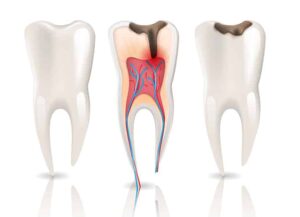
Root canal treatment is needed when the pulp — the nerve and blood supply inside the tooth — is irreversibly inflamed or infected. The usual route there is decay that reached the pulp, or a crack that let bacteria in. See how does tooth decay develop?, the stages of dental decay and can you reverse tooth decay, and do I need a filling? — the last of which describes the window in which none of this is necessary.
What the pain does and does not tell you is covered in what are the causes of toothache and what are their symptoms?, I have a toothache — what could be the cause? and tooth pain and toothaches. If it has flared up out of hours, emergency dentistry and what is considered a dental emergency?
What an endodontist is
Endodontists are dentists who have trained for a further three years after their basic dental training to specialise in root canal treatment. See Endodontists and Dentists & Registered Specialists.
That advanced training includes the use of specialised equipment — which is the point of this article. See Technology.
Root canal treatment is within the scope of general dentistry, and many general dentists perform it well. What the specialist training and equipment add is the ability to handle the cases where the anatomy is complex, unusual, or hidden. Endodontist vs dentist for root canal sets out which cases fall where.
When treatment does fail, the most common reason is infection — typically because bacteria remained somewhere in the canal system that was not found and cleaned. At that point patients are commonly referred to an endodontist for re-treatment, or advised to have the tooth extracted. See What is a tooth abscess? and Second Opinions & Corrective Dentistry.
Re-treating a root canal is considerably harder than doing it once, which is the practical argument for getting the difficult cases to the right hands the first time. The measured gap is modest: in a Taiwanese registry study of more than three million teeth, five-year survival was 90.9% for teeth root-treated for the first time and 88.4% for retreated teeth.
If the tooth cannot be kept, what follows is set out in what are the replacement options for missing teeth? and implant versus bridge for a single tooth replacement — both of which cost more than keeping the tooth.
The microscope
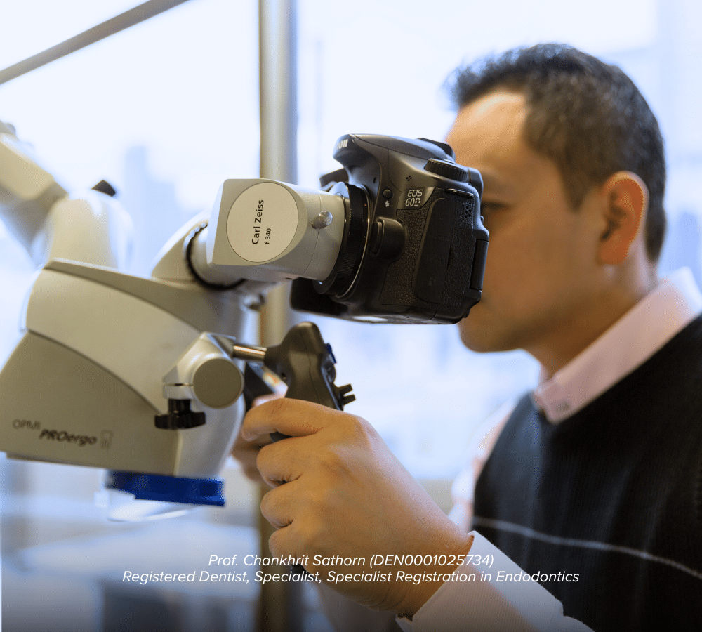
A key development in the success of root canal treatment is the use of high-powered microscopes.
Pioneered in the late 1990s, the operating dental microscope lets the operator see inside a tooth and its root canal system at magnification of up to 20 times, with added lighting.
Both halves matter. Magnification without illumination is of limited use inside a tooth, because you are looking into a narrow, dark chamber. The microscope delivers coaxial light down the same axis you are viewing along.
Why it changes outcomes
Teeth often have extra roots or canals that cannot be seen with the naked eye or with loupes (magnifying glasses).
This is the decisive one. An unfound canal is an uncleaned canal, and an uncleaned canal holds bacteria. Root canal treatment fails not because the found canals were treated badly but because a canal was never found at all. Certain teeth commonly have an additional canal that is small, off-centre and easy to miss — and the difference between finding it and not finding it is largely optical.
What “extra anatomy” actually means. The British Dental Journal review lists the variants an operator is hunting for: a second canal in the mesio-buccal root of an upper molar, a second lingual canal in lower incisors, three-rooted premolars, an additional root on a lower molar (radix entomolaris), and complexities such as C-shaped canals, dens invaginatus, an isthmus running between two canals, apical deltas and lateral canals — along with canals of extreme curvature. That review describes untreated anatomy housing persistent endodontic infection as one of the widely accepted causes of treatment failure.
Crowns or large fillings often make it difficult to see inside a tooth, and the microscope's light is needed to see the floor of these teeth — the surface where canal openings are located. See Dental Crowns and Tooth Fillings.
Fractures and other abnormal anatomy can be detected and treated early, rather than discovered after the fact, when treatment has already failed. A vertical root fracture found before treatment saves a patient the cost and time of a root canal on a tooth that could not have been saved. See Chipped and Cracked Teeth, why does a cracked tooth hurt so much? and how will my cracked tooth be treated?
What the microscope cannot do is see through bone, which is what radiographs are for — how safe are dental x-rays?
The microscope is not the only instrument that matters. Rotary nickel-titanium files, apex locators and activated irrigation all contribute — and each has its own failure mode, such as the risk of broken files during root canal treatment.
What else the outcome turns on
How big the lesion was before treatment started
The presence of a periapical lesion is a negative prognostic factor, and size matters in a measurable way. The British Dental Journal review quotes a prospective study which concluded that “the odds of success of treatment were found to decrease by 14% for every 1 mm increase in diameter of the preoperative lesion”.
The 21-year cohort found the same thing from the other direction: a pre-operative periapical radiolucency was significantly associated with the tooth eventually being extracted (odds ratio 1.87; 95% CI 1.07 to 3.28; p = 0.028).
This is the clearest argument available for not waiting. The lesion does not shrink while you think about it.
Whether the tooth is cracked
The presence, or inferred presence, of a crack is a negative prognostic factor for survival — a crack is both a route for bacteria and a structural weakness. The review notes a useful sign: a localised, narrow, deep pocket beside an otherwise healthy tooth is often considered characteristic of a crack that has reached the root surface. If a hygienist has mentioned an isolated deep pocket next to one tooth, raise it specifically — see Dental Cleans & Hygienists and Periodontists.
Whether the tooth was properly isolated
Treatment is carried out under a rubber dam — a sheet that isolates the tooth from the saliva that carries the bacteria being cleaned out. In large registry studies from Taiwan and Korea, recorded use of a rubber dam significantly and positively affected the five-year tooth survival rate.
A separated instrument is not automatically a failure
Files occasionally fracture inside a canal. The British Dental Journal review is specific: if the instrument can be removed or bypassed, there is no negative effect on treatment outcome. If it cannot be, and a periapical lesion is present, the infection at the root tip becomes difficult to reach and the outcome is less predictable. See the risk of broken files during root canal treatment.
After the root canal
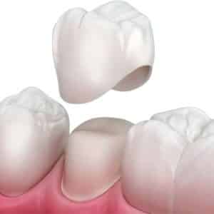
A root-treated back tooth is usually crowned, because removing the pulp and the access cavity leaves it more liable to split. That is a second appointment and a second fee, and it should be in the plan and the quote from the outset — see what types of dental crowns are available?, how much does a dental crown cost in Melbourne?, the price guide and understanding your treatment.
The restoration is not an optional extra
A systematic review cited in the British Dental Journal paper concluded that the coronal seal was as important as the quality of the root canal treatment itself in determining success, and there is evidence of an increased failure rate where teeth are left in temporary restorations. Timing has been measured too: in a retrospective study, posterior root-filled teeth that received a crown four months after treatment were extracted at three times the rate of those crowned within four months.
The 21-year cohort adds detail about what the tooth is restored with:
- a cast metal post was associated with extraction (OR 2.14; 95% CI 1.14 to 4.01; p = 0.018)
- a fibre post was protective (OR 0.47; 95% CI 0.24 to 0.91; p = 0.026)
- probing pocket depth of 5 mm or less was protective (OR 0.68; 95% CI 0.54 to 0.86; p = 0.001) — that is, healthy gums around the tooth
- wearing a night guard was protective (OR 0.34; 95% CI 0.13 to 0.86; p = 0.023)
If you grind your teeth, say so, because a root-treated, crowned tooth takes that load without the early warning a live nerve would give — night time tooth grinding and clenching, what is bruxism and how is it managed? and how can a night guard be used to treat TMD?
The tooth still decays at the margin like any other, so the ordinary routine still applies — what is the ideal daily routine for oral hygiene?, is flossing really that important? and Dental Cleans & Hygienists.
What actually takes these teeth out
Of the 598 teeth followed, 87 (14.5%) were extracted, after a mean of 14.1 years. The recorded reasons, as a share of the teeth that were lost, were:
- periodontal disease progression — 40.2%
- vertical root fracture — 33.3%
- crown fracture — 15%
- caries — 11.5%
- endodontic inflammation — 0%
The authors singled that last line out: in that cohort, no tooth was extracted because of endodontic inflammation. The teeth that were lost were lost to gum disease, to the tooth splitting, and to new decay.
That is a finding about one group of patients in one practice over 21 years. It is not a promise about any individual tooth, and it cannot be read as one. But it points somewhere practical: once a canal has been found, cleaned and sealed, the things most likely to cost you that tooth afterwards are the ordinary ones — your gums, your bite and your brushing. Which is an argument for regular maintenance rather than for relief.
What this means for you
Advances in technology and training have for some time allowed endodontists to achieve high success rates.
Contrary to outdated perceptions, treatment does not always require multiple visits, and modern techniques and anaesthesia mean it is generally comparable to having a filling placed. For anyone whose reluctance is about the dental chair rather than the tooth, see Dental Anxiety, how can I ease my anxiety about visiting the dentist?, how injections are made more comfortable and Sleep Dentistry.
A reasonable question to ask before root canal treatment: will a microscope be used? It is not a marketing question. It bears directly on whether every canal in the tooth gets found and cleaned, which is the single largest determinant of whether the treatment holds.
Common questions
Does it hurt? And why does nobody answer that with a number?
The second half of that question has a genuinely interesting answer, and it is more useful than a reassurance.
The British Dental Journal review of endodontic outcomes is candid that the profession measures this badly. It records “the lack of good-quality evidence in this important area of understanding the patient's perception of endodontic outcome,” and attributes it to “the poor validity of the measurement tools, making conclusions difficult to draw.” In other words: radiographic healing has been studied exhaustively and can be quoted to two decimal places, while the patient's actual experience has not been measured well enough to quote at all.
So the honest position is this. Discomfort during treatment is managed with local anaesthetic, and the common modern experience is described above as generally comparable to having a filling placed. Some tenderness on biting for a few days afterwards is ordinary, and it settles. What nobody can honestly give you is a published figure for how it will feel, because that figure does not exist in usable form.
What you can do is ask specifically: what will be done if I feel something during the appointment, how do we stop, and what should I expect for the next two or three days. A clinician who answers those three in detail is telling you more than any statistic would.
One thing worth separating out: a tooth that is already acutely inflamed can be harder to numb than a quiet one, which is an argument for dealing with it earlier rather than waiting for it to flare.
Should I just have it out instead? It would be cheaper today.
It would be, and the question deserves a straight comparison rather than a lecture.
The case for keeping the tooth is in the figures above: cumulative success around 93 per cent at ten years in the 21-year cohort, and survival of the tooth in function around 97 per cent at ten years. Against that, an extraction is permanent, and the replacement — a denture, a bridge or an implant — costs more than the root canal and the crown together, carries its own maintenance, and, in the case of an implant, needs the bone that removing the tooth begins to lose.
The case for extraction is stronger than most patients realise in one specific circumstance: when there is not enough sound tooth left to restore. The British Dental Journal review cites a study of root-canal-retreated posterior teeth in which, where less than 29.5 per cent of tooth structure remained, the rate of extraction was three times higher than in teeth with more than 29.5 per cent remaining. Restorability, not the canal, is often the deciding variable — and it is a judgement that can be made before you commit, not after.
So the questions to put are: how much sound tooth is left, is this tooth restorable, is there a crack, and what would the replacement cost and involve. If the answer to the second is no, paying for a root canal first is the expensive route to the same extraction.
Will I need a crown, what kind, and how soon?
Usually yes for a back tooth, and the timing is the part with a measured answer.
How soon. As noted above, a retrospective study found posterior root-filled teeth crowned four months after treatment were extracted at three times the rate of those crowned within four months. The guidance drawn from that is to provide the definitive restoration as soon as possible once the tooth is settled, rather than leaving it in a temporary.
Whether. The decision to provide cuspal coverage — a restoration that covers the biting surface, rather than a filling in the access cavity — is made case by case; both the British Endodontic Society's guide and the European Society of Endodontology's position statement say so explicitly. The factors named are how much tooth structure remains, tooth position, the adjacent contacts and the forces on it. Loss of the proximal walls is described as a strong indication that cuspal coverage is needed, and loss of a proximal contact, or being the terminal tooth in the arch or a second molar, is associated with a higher risk of failure.
What kind. A systematic review cited in the same paper gives five-year single-crown survival rates by material: metal-ceramic 94.7 per cent, leucite or lithium-disilicate reinforced glass ceramic 96.6 per cent, and densely sintered zirconia 92.1 per cent. A separate prospective study reported 92.3 per cent at five years for gold crowns. Those are close enough that the choice usually turns on appearance, the space available and cost rather than on durability — and the guidance is to conserve as much sound tooth as possible, using an onlay rather than a full crown where that is appropriate.
What types of dental crowns are available? covers the materials, and published fees are in the price guide.
How will I know it worked?
Not by how it feels, which is the point most people miss.
The definition quoted at the top of this page has two halves: no symptoms or clinical signs of disease, and a radiograph showing an intact apical periodontal ligament space with any previous radiolucency resolved. Bone healing takes time — it is assessed over months to years rather than at the next appointment — so the follow-up radiograph is the instrument that answers the question, not the absence of pain.
The reason that matters is the figure quoted above: around 40 per cent of root-filled teeth show apical periodontitis when populations are surveyed cross-sectionally. Most of those people have no symptoms at all. A tooth that feels fine and a tooth that is healing are not the same finding.
Practically: ask what the review interval is and attend it, and mention the tooth at ordinary check-ups so it is deliberately looked at rather than passed over. If something does change — a swelling, a gum boil near the root, discomfort on biting that arrives months later, or a tooth that starts to feel loose — that is a reason to be seen rather than to wait for the scheduled review.
Related reading
- Root canal treatment: who and what is involved?
- Everything you need to know about root canal treatment
- Why are dental abscesses so painful?
- Can a dental abscess affect your general health?
- How do I prevent dental decay?
- How can I relieve a toothache?
Practical details
Smile Solutions has registered specialist endodontists on site, with operating microscopes. The clinicians are listed on Our Team.
Smile Solutions, Level 1, 220 Collins Street, Manchester Unity Building, Melbourne VIC 3000 — getting here. Phone 13 13 96, or theteam@smilesolutions.com.au. Monday–Friday 8.00am–6.00pm, Saturday 8.30am–1.30pm, Sunday by appointment. Full details on Contact Us.
Specialist registration can be verified on the AHPRA register, or by calling 1300 419 495.
Published 8 April 2018. Success rates are figures from published research, not guarantees; outcomes vary between individuals and all treatment carries risks that should be discussed with your clinician. Research figures quoted here come from independent peer-reviewed sources and describe study populations, not individual patients. General information only; it does not replace advice from your treating practitioner.
Smile Solutions trades under ABN 28 193 514 103.
Images on This Page
-
https://www.facebook.com/tr?id=800557773428361&ev=PageView&noscript=1
(no alt text)
-
https://www.smilesolutions.com.au/wp-content/uploads/2025/10/calendar_month.svg
(no alt text)
-
https://www.smilesolutions.com.au/wp-content/themes/arcadian-wordpress-theme/assets/images/smile_solutions_logo_white.png
Smile Solutions Logo
-
https://www.smilesolutions.com.au/wp-content/themes/arcadian-wordpress-theme/assets/images/smile_solutions_logo_purple.png
Smile Solutions Logo
-
https://www.smilesolutions.com.au/wp-content/uploads/2023/11/SS-Tooth-1-1-150x150.png
Smile Solutions
-
https://www.smilesolutions.com.au/wp-content/uploads/2025/09/Root-Canal-Therapy-Endodontist-Smile-Solutions.png
Specialist Endodontist using a dental microscope
-
https://www.smilesolutions.com.au/wp-content/uploads/2025/09/Root-Canal-2-300x217.jpg
Root Canal
-
https://www.smilesolutions.com.au/wp-content/uploads/2026/08/Mouth-Breathing-300x270.jpg
Mouth Breathing
-
https://www.smilesolutions.com.au/wp-content/uploads/2025/09/Dental-Crown-at-Smile-Solutions-300x300.jpg
Dental Crown at Smile Solutions
-
https://www.smilesolutions.com.au/wp-content/themes/arcadian-wordpress-theme/assets/images/family_owned_bussiness.png
Family Owned Business
-
https://www.smilesolutions.com.au/wp-content/themes/arcadian-wordpress-theme/assets/icons/icon--phone.svg
Smile Solutions Contact
-
https://www.facebook.com/tr? id=800557773428361&ev=PageView&noscript=1
(no alt text)
-
https://bat.bing.com/action/0?ti=25148060&tm=gtm002&Ver=2&mid=d326be32-2be5-4907-ac21-625151d08678&bo=1&sid=1ed17a80ab2711f196506bcf7bb7b307&vid=1ed3e530ab2711f1b68e3f9bc05b191c&vids=1&msclkid=N&pi=918639831&lg=en-US&sw=800&sh=600&sc=24&nwd=1&tl=Why%20is%20the%20microscope%20so%20crucial%20in%20endodontic%20treatment%20by%20a%20specialist%3F%20-%20Smile%20Solutions&p=https%3A%2F%2Fwww.smilesolutions.com.au%2Fdental-articles%2Farticle%2Fmicroscope-so-crucial-in-endodontic-treatment%2F&r=&evt=pageLoad&sv=2&cdb=AQAQ&rn=961925
(no alt text)