How safe are dental X-rays?
The short answer, with the number attached
A single intraoral dental X-ray — a bitewing or a periapical — delivers a typical effective dose of 1 to 8 microsieverts (μSv), which is 0.001 to 0.008 millisieverts (mSv). The annual dose limit ARPANSA sets for a member of the public is 1 mSv.
That is the comparison worth holding onto: an intraoral film is roughly 0.1 to 0.8 per cent of a year's public dose limit. A panoramic image (OPG) is larger, and cone beam CT larger again; the dose figures are the International Atomic Energy Agency's, and the full table is further down this page.
One caveat on that comparison: dose limits do not apply to your own dental imaging. ARPANSA's limits govern occupational and public exposure, while a radiograph taken for your diagnosis is justified case by case instead. The 1 mSv figure is a yardstick for scale, nothing more.
This does not mean the dose is zero, and it should not be presented that way. There is a known relationship between radiation dose and cancer risk. What it means is that the dose from dental imaging is very small, and that the decision to take an X-ray is a judgement about whether the diagnostic information is worth that small exposure. The companion article How safe are dental X-rays, and when do they become unsafe? takes the second half of that question further.
Why an X-ray is taken at all
This is the part most people skip past, and it is the part that justifies the exposure.
A visual examination cannot see everything. Specifically, it cannot assess:
Decay between teeth (interproximal decay). The contact point between two adjacent teeth is not visible to the eye or a probe. Decay can be well established there and completely invisible on visual examination. The stages of dental decay explains how far it can progress silently, and can you reverse tooth decay? why finding it early changes the treatment.
The level of the alveolar bone. The bone that holds the teeth in their sockets sits below the gum. Bone loss from periodontal disease cannot be measured by looking — it has to be imaged. See also what is gum disease? and when do you need deeper cleaning?
Suspicious lesions in the bone and roots. Cysts, abscesses at root tips, resorption, unerupted or impacted teeth, and other pathology inside bone are simply not visible from the outside. An abscess is not a contained problem — can a dental abscess affect your general health?
The practical consequence: a dentist working without X-rays is diagnosing with a substantial part of the picture missing. Decay found early on a bitewing is a small filling. The same decay found later, when it has become visible or symptomatic, may be a root canal treatment and a crown. The difference in cost between those two is set out in the price guide, and the difference in what is involved in everything you need to know about root canal treatment.
That is the trade-off: a very small radiation dose against the risk of missing disease that is treatable while it is small.
The three types you will encounter
Bitewing X-ray
So named because you bite down on a tab holding the film or sensor in place. Shows the crowns of the upper and lower back teeth together, plus the bone level between them.
Its main job is finding decay between teeth, and monitoring bone levels over time. This is the routine one — the image most often taken as part of a check-up.
Intraoral periapical X-ray
“Peri-apical” means around the apex — the tip of the root. Shows the entire tooth from crown to root tip, plus the surrounding bone.
Taken when there is a specific concern about a particular tooth: pain, an abscess, before or after root canal treatment, or to check a root fracture. Where a tooth is being retreated, the imaging is part of what the endodontist works from — see why the microscope is so crucial in endodontic treatment.
OPG (orthopantomogram)
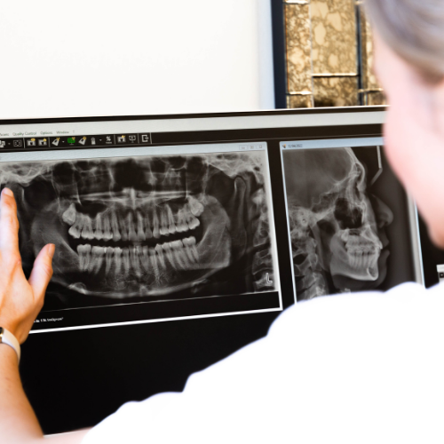
A panoramic image taken with the machine rotating around your head. Shows all the teeth, both jaws, the sinuses and the jaw joints in one picture.
Used for the overall view: wisdom teeth position, unerupted teeth, jaw pathology, orthodontic planning, and implant assessment. It gives less fine detail than an intraoral film, so it complements rather than replaces bitewings. Implant planning in particular often needs the three-dimensional view as well, especially where bone grafting may be required — see what do I need to know about dental implants?
Orthodontic assessment usually adds a fourth image, a cephalometric view of the side of the head, used to measure how the jaws sit relative to one another — see when should I take my child to see an orthodontist?
What digital imaging changed
Advances in digital radiographic technique have significantly reduced the radiation dose compared with the film-based systems they replaced. The current equipment is listed under technology.
Digital sensors are more sensitive than film, so less radiation is needed to produce a usable image. There is no chemical processing, which means no retakes because a film was developed poorly — and every avoided retake is an avoided dose.
This is the main reason older figures for dental X-ray exposure, which you will still find quoted online, overstate the dose from a modern digital system. Dental myths exposed deals with several other claims of that vintage.
Putting the number in context
| Dose | |
|---|---|
| Single intraoral dental X-ray (IAEA) | 1–8 μSv = 0.001–0.008 mSv |
| Annual public dose limit (ARPANSA) | 1 mSv |
| Annual occupational dose limit (ARPANSA) | 20 mSv per year averaged over 5 years, and not more than 50 mSv in any one year |
The ratio is the answer to the question: an intraoral dental X-ray is roughly 0.1 to 0.8 per cent of the annual public dose limit, and less than one day of the 1.7 mSv of natural background radiation ARPANSA says the average Australian receives each year.
The occupational figures are routinely misstated, so they are worth quoting exactly — “20 millisieverts (mSv) per year averaged over 5 years” and “not more than 50 mSv received in any one year for effective (whole body) dose”. The 50 mSv is a single-year ceiling, not the annual limit, and quoting it as the annual limit overstates it two and a half times, which makes any dental comparison look more favourable than the correct figure does.
What the independent source puts the dose at
The International Atomic Energy Agency publishes typical effective doses for dental procedures — effective dose being the quantity that weights each organ by how sensitive it is to radiation:
| Examination | Typical effective dose (IAEA) |
|---|---|
| Intraoral — bitewing or periapical | 1–8 μSv |
| Panoramic (OPG) | 4–30 μSv |
| Cephalometric | 2–3 μSv |
| CBCT, small or medium scanning volume | 50 μSv or below |
| CBCT, large volume | around 100 μSv |
One thousand microsieverts (μSv) make one millisievert, so the whole table sits between 0.001 and 0.1 mSv. Three things follow.
Which examination you had matters more than which machine took it. A panoramic image covers both jaws and can be several times the dose of a single intraoral film, so a number quoted for “a dental X-ray” without saying which one is close to meaningless. Before concluding that two published figures contradict each other — including on this site — check whether they describe the same examination.
The IAEA frames the dose against natural background rather than an occupational limit. Intraoral and cephalometric procedures come to less than one day of natural background radiation; panoramic procedures, even at the top of their range, to a few days of it, which the IAEA describes as similar to a chest radiograph.
Cone beam CT is a different conversation. The IAEA notes CBCT can run tens or even hundreds of microsieverts above conventional techniques, and that typical ranges are likely to change as equipment improves — so an older quoted figure may not describe the machine in front of you. If a CBCT is proposed, ask what field of view is being used.
One IAEA caution usually gets lost: effective dose exists to compare modalities and techniques, and is not intended to be applied to an individual person. It does not calculate your risk.
Is there an Australian benchmark for dental doses? No
This changes what “within the guidelines” can honestly mean.
ARPANSA, the Australian Radiation Protection and Nuclear Safety Agency, runs the National Diagnostic Reference Level Service. A diagnostic reference level is not a regulatory limit — it is a benchmark set at the 75th percentile of a national survey, and a facility above it is expected to review its protocols, not to stop imaging.
ARPANSA has concentrated on the procedures carrying the highest dose burden nationally, publishing reference levels for multi-detector CT in adults and children, general nuclear medicine and PET, the CT component of SPECT/CT and PET/CT, and diagnostic coronary angiography. It states plainly that there are no Australian diagnostic reference levels for planar radiography, dental X-ray, cone beam CT or mammography.
So if you are told a dental dose is “below the Australian reference level”, there is no such level to be below. The honest version is that dental doses are small enough not to have been prioritised for national benchmarking — reassuring about the size of the dose, and a good reason to ask the indication question instead.
Deterministic and stochastic: the distinction behind “is it safe”
Almost every confused argument about dental X-rays comes from mixing up two different kinds of effect.
Deterministic effects — tissue reactions — result from killing cells and only occur above a dose threshold. A peer-reviewed review of intraoral radiography in children states that conventional dental imaging never causes them.
Stochastic effects work by damaging DNA. They can follow very low exposures, no threshold dose has been demonstrated, and the risk extends over a lifetime. That is why the honest answer is never “X-rays are harmless”, and why every image needs a reason.
The same review puts the working principle bluntly: there is a health risk with every X-ray, the duty is to keep it as low as possible, and no X-ray necessary for appropriate dental care should be withheld. Declining a justified radiograph is not the cautious choice — it trades a very small quantified risk for an unquantified one.
What the image actually buys, measured
The justification for imaging is usually asserted rather than measured. It has been measured: a peer-reviewed review reports that visual examination supported by intraoral or bitewing radiographs detected up to 50 per cent more decay lesions on the surfaces between back teeth than visual examination alone, in both baby and adult teeth.
That is the diagnostic gap the exposure is traded against. The value of closing it early is not earlier drilling — it is the option of not drilling, monitoring and intensifying prevention while the lesion is still confined to enamel. See can you reverse tooth decay, and do I need a filling?, the benefits of fluoride and dental cleans and hygienists.
Children, and why the standard is stricter
Children are more vulnerable to radiation than adults, for specific reasons rather than precautionary ones: a higher rate of cell division, a higher proportion of water in the tissues, radiation-sensitive organs close to the field — the lens of the eye, the thyroid and pituitary glands, the oral cavity — and a longer expected lifetime in which a stochastic effect could appear.
Routine X-ray screening of children is no longer recommended. Taking radiographs at fixed intervals according to decay risk has been superseded; European and American paediatric dentistry bodies both now require individualised, patient-specific justification for each image, with the American guidance stating that timing should be based on the child’s individual circumstances and not on their age.
The guiding principle has also shifted from ALARA to ALADA — from “as low as reasonably achievable” to as low as diagnostically acceptable, indication-oriented and patient-specific. That is not cosmetic: a dose too small to give a diagnostic image is no saving, because the image gets repeated.
In practice the commonest reasons children are imaged are dental trauma, decay diagnosis and assessment around the root tip — see children’s dentistry, should your child see a specialist paediatric dentist? and, for a knocked or broken tooth, emergency dentistry.
For intraoral imaging the established dose-reduction measures are worth knowing by name, because you can ask about them: rectangular collimation (shaping the beam to the sensor rather than sending a round beam wider than the image needs), fast image receptors, thyroid shielding, and exposure settings optimised for the task rather than left at a default.
Common questions
Safety is not only about the size of the dose. It is also about whether the image was necessary — which is the part you can actually ask about.
Why is this X-ray needed?
There should be a specific diagnostic reason, and “it is time for your X-rays” is not one. Reasonable answers sound like a question the image will settle: whether the shadow between two teeth is decay, how far the bone has been lost, why that tooth is tender, where an unerupted tooth is sitting.
This is not a sceptical question, it is the standard the profession applies to itself. The IAEA lists justification as one of the core radiation-protection principles in dentistry, and the peer-reviewed review of paediatric intraoral imaging states the position without hedging: “There is a certain health risk associated with every X-ray. It is our duty to keep this risk as low as possible.” The same passage adds the other half, which matters just as much — no X-ray necessary for appropriate dental care should be withheld. Justification runs both ways.
Understanding your treatment covers how findings and the plan that follows should be explained to you.
What would you do differently depending on what it shows?
The sharper version of the previous question, and the one that is hardest to answer vaguely. A justified radiograph changes a decision — between filling and monitoring, between a general dentist and a specialist, between extracting a wisdom tooth and leaving it.
It is also the test current paediatric guidance applies. The shift away from fixed-interval screening in children, and from ALARA to ALADA — as low as diagnostically acceptable — is a shift towards asking what the image will be used for before it is taken. American paediatric guidance is explicit that timing should follow the child’s individual circumstances rather than their age.
If the honest answer is “probably nothing, but it is good to have a record”, that is worth hearing too. It may still be a reasonable image to take. It is simply a different reason from the one you were given.
How often do I need them?
There is no universal interval, and anyone quoting one is describing a scheduling habit rather than a clinical standard. The interval should follow your own decay risk and periodontal status — someone with no new decay in a decade and stable bone levels is not on the same schedule as someone with active lesions, a dry mouth or deepening pockets.
Two things support treating this as individual rather than fixed. First, routine fixed-interval radiographic screening of children has been superseded by patient-specific justification, in both the European and American paediatric guidance. Second, ARPANSA publishes no Australian diagnostic reference level for dental X-ray at all — it has set reference levels for multi-detector CT, nuclear medicine and PET, the CT component of SPECT/CT and PET/CT, and coronary angiography, and states that none exist for planar radiography, dental X-ray, cone beam CT or mammography. So there is no national number to be measured against, in either direction.
Ask what interval you have been put on, what would shorten it, and what would lengthen it. The same logic governs how often you should go to the dentist.
Are previous X-rays available?
Ask before anything is taken. If recent images exist from another practice, they can often be obtained rather than repeated — and an avoided retake is an avoided dose, which is the same logic that makes digital sensors preferable to film.
Older images are also worth more than new ones for some purposes. Bone levels and slow-growing decay are read from change, so a five-year-old bitewing next to today’s is more informative than today’s alone. Request copies when you leave a practice, and keep them; you own the information even where the practice owns the record.
This is one practical advantage of continuity — see is it important to have a family dentist? — and it applies equally when you are getting a second opinion.
I am pregnant, or might be. Should I have a dental X-ray?
Tell your dentist first, before anything is set up, and expect the answer to depend on whether the image is urgent.
Dental X-rays are generally considered low risk in pregnancy with appropriate shielding, and the doses involved are the ones in the table above — an intraoral film at 1 to 8 μSv, less than a day of natural background radiation. In practice, non-urgent imaging is commonly deferred anyway, and that is a reasonable default rather than an alarmed one. What matters is that the decision is made knowingly: if there is a tooth causing pain or a suspected infection, delaying the image can be the greater risk, and untreated dental infection in pregnancy is not a small matter.
The IAEA maintains specific radiation-protection guidance for pregnant patients in dentistry, so this is a question your clinician should be able to answer from a source rather than from instinct. See is it safe to visit the dentist during pregnancy? and oral health care while pregnant.
Can I say no?
Yes. Consent applies to imaging as it does to treatment, and you are entitled to decline.
What you should also hear is what declining costs, because it is not a neutral choice. Visual examination alone misses decay that imaging finds: the peer-reviewed review cited above reports that visual examination supported by intraoral or bitewing radiographs detected up to 50 per cent more decay lesions on the surfaces between back teeth than visual examination alone, in both baby and adult teeth. Bone loss cannot be measured by looking at all.
So the honest framing is the one the same review uses — the duty is to keep the risk as low as possible, and no X-ray necessary for appropriate dental care should be withheld. Declining a justified radiograph trades a very small quantified risk for an unquantified one. If you want to decline, say so and ask what will be missed and what the plan is instead; a reasonable clinician will document that and work with it.
Should I be given a lead apron or a thyroid collar?
Worth asking, and worth knowing that the two are not on the same footing in the evidence we can point to.
Thyroid shielding is named explicitly in the peer-reviewed review of intraoral radiography, alongside rectangular collimation and fast image receptors, as a protective device that reduces radiation exposure in intraoral imaging. The thyroid is one of the radiation-sensitive organs close to the dental field, which is why it is singled out — particularly in children.
The abdominal lead apron is a different case, and we are not going to overstate it: no source in our own reference set addresses whether a lead apron adds protection in dental imaging. International practice has moved in different directions on this, and it is a fair question to put to your clinician — ask what shielding they use, for which examinations, and why. What we can say is that the collimation and receptor choices matter, because those reduce the dose at source rather than catching it afterwards.
I have had CT scans this year. Does my dental imaging add to that?
It adds to your cumulative exposure, but far less than the question usually assumes, and the arithmetic does not work the way people expect.
On scale: the entire dental table above sits between 0.001 and 0.1 mSv per examination. A medical multi-detector CT is in a different order of magnitude, which is precisely why ARPANSA has set national diagnostic reference levels for CT and not for dental X-ray.
On the arithmetic, two cautions from the sources themselves. The IAEA is explicit that effective dose and its associated risk “should not be applied to individuals” — it exists to compare modalities and techniques, not to compute a personal total. And the ARPANSA dose limits quoted above govern occupational and public exposure; they do not apply to your own diagnostic imaging, which is justified case by case instead. So there is no running total you are at risk of exceeding, and no threshold at which a dentist should refuse a justified radiograph because of a CT you had in March.
What is genuinely useful is to tell your dentist what imaging you have had and when — not to keep a score, but because an existing image may answer the question and save a new one.
Does this article replace an individual dental assessment?
No. It provides general information. Symptoms, suitability, risks and treatment choices depend on examination and, where indicated, imaging or other records from an appropriately registered practitioner.
Related reading
- How safe are dental X-rays and when do they become unsafe?
- The benefits of holistic dentistry — which covers the digital-dose question from the materials side
- What is holistic dentistry?
- Oral cancer: how your dentist can help with early detection
- Why would I need to see a dental specialist?
- 5 questions you’ve always wanted to ask your dentist
- General dentistry and our team
Where the external figures come from
- Effective doses by examination type, and the comparison with natural background: International Atomic Energy Agency, Radiation doses in dental radiology.
- Public and occupational dose limits, and average natural background radiation in Australia: ARPANSA, Information for workers and Occupational exposure: Medical workers, the limits being those prescribed in the Code for Radiation Protection in Planned Exposure Situations (RPS C-1).
- Diagnostic reference levels, and their absence for dental X-ray and cone beam CT: ARPANSA, National Diagnostic Reference Level Service.
- Deterministic and stochastic effects, paediatric radiosensitivity, ALADA, thyroid shielding and the caries-detection comparison: peer-reviewed study of intraoral radiograph exposure in children and adolescents, indexed on PubMed Central.
Every dose figure on this page is taken from a published source and attributed to it. An earlier version gave 0.02 mSv for a dental X-ray and 50.0 mSv as the Australian annual occupational limit, both flagged as Smile Solutions’ own approximations. The first sat above the IAEA intraoral range quoted on this same page; the second is a single-year ceiling. Both have been replaced.
Practical details
Smile Solutions, Level 1, 220 Collins Street, Manchester Unity Building, Melbourne VIC 3000 — see how to find us. Phone 13 13 96, or theteam@smilesolutions.com.au; full details on the contact page. Monday–Friday 8.00am–6.00pm, Saturday 8.30am–1.30pm, Sunday by appointment.
Published 8 January 2024. Dose figures are typical published ranges and vary with equipment and technique. General information only; it does not replace advice from your treating practitioner.
Smile Solutions trades under ABN 28 193 514 103.
Images on This Page
-
https://www.facebook.com/tr?id=800557773428361&ev=PageView&noscript=1
(no alt text)
-
https://www.smilesolutions.com.au/wp-content/uploads/2025/10/calendar_month.svg
(no alt text)
-
https://www.smilesolutions.com.au/wp-content/themes/arcadian-wordpress-theme/assets/images/smile_solutions_logo_white.png
Smile Solutions Logo
-
https://www.smilesolutions.com.au/wp-content/themes/arcadian-wordpress-theme/assets/images/smile_solutions_logo_purple.png
Smile Solutions Logo
-
https://www.smilesolutions.com.au/wp-content/uploads/2023/11/SS-Tooth-1-1-150x150.png
Smile Solutions
-
https://www.smilesolutions.com.au/wp-content/uploads/2025/09/Wisdom-Teeth-Smile-Solutions.png
Smile Solutions Dentist Melbourne
-
https://www.smilesolutions.com.au/wp-content/uploads/2025/09/Root-Canal-2-300x217.jpg
Root Canal
-
https://www.smilesolutions.com.au/wp-content/uploads/2026/08/Mouth-Breathing-300x270.jpg
Mouth Breathing
-
https://www.smilesolutions.com.au/wp-content/uploads/2025/09/Dental-Crown-at-Smile-Solutions-300x300.jpg
Dental Crown at Smile Solutions
-
https://www.smilesolutions.com.au/wp-content/themes/arcadian-wordpress-theme/assets/images/family_owned_bussiness.png
Family Owned Business
-
https://www.smilesolutions.com.au/wp-content/themes/arcadian-wordpress-theme/assets/icons/icon--phone.svg
Smile Solutions Contact
-
https://www.facebook.com/tr? id=800557773428361&ev=PageView&noscript=1
(no alt text)
-
https://bat.bing.com/action/0?ti=25148060&tm=gtm002&Ver=2&mid=ec60d777-32d0-4415-b30c-cfea9a05dc1a&bo=1&sid=de0dd3b0ab2711f1b8f1c5048ad66ebb&vid=de0e0030ab2711f1940375526045ea7d&vids=1&msclkid=N&pi=918639831&lg=en-US&sw=800&sh=600&sc=24&nwd=1&tl=How%20safe%20are%20dental%20x-rays%C2%A0%20-%20Smile%20Solutions&p=https%3A%2F%2Fwww.smilesolutions.com.au%2Fdental-articles%2Farticle%2Fhow-safe-are-dental-x-rays%2F&r=&evt=pageLoad&sv=2&cdb=AQAQ&rn=429512
(no alt text)