Why jaw pain treatment so often goes in circles
There is a reason patients with jaw problems feel like they are going round in circles.
They see a dentist who says they grind and makes a night guard. A physiotherapist who treats their neck. A GP who prescribes anti-inflammatories. Each practitioner addresses one piece of the puzzle based on their own examination. None has the full picture.
The fundamental challenge with diagnosing temporomandibular disorders (TMD) is that clinical examination alone — even by an experienced practitioner — has real limits: If the terminology is unfamiliar, start with TMD vs TMJ vs bruxism: understanding what's actually wrong with your jaw and what are the most common symptoms of TMD?
- You can palpate muscles for tenderness, but you cannot feel which ones are overactive and by how much
- You can check the bite with articulating paper, but you cannot see the timing and magnitude of forces across every tooth simultaneously
- You can take a standard X-ray, but you cannot visualise the three-dimensional anatomy of the joint, the airway, or the disc
Where traditional diagnosis falls short
History, clinical examination and standard radiography remain the foundation of every assessment. But they have documented limitations — and it is worth being precise about where those limits actually lie.
Palpation depends on protocol
When a clinician presses on a muscle and asks whether it hurts, the answer depends on the patient's pain threshold, their anxiety level, the pressure applied — which varies between clinicians — and whether the muscle happens to be tender at that moment.
Standardising that pressure changes the picture considerably. The Diagnostic Criteria for Temporomandibular Disorders (DC/TMD), published in the Journal of Oral & Facial Pain and Headache in 2014, specify palpation at 1 kg of pressure for 2 seconds, extended to 5 seconds to distinguish the types of muscle pain — and report that, used that way, inter-examiner reliability for pain-related TMD is "excellent (kappa ≥ 0.85)", with sensitivity of at least 0.86 and specificity of at least 0.98. So careful, protocol-driven examination of muscle pain is reliable, and this page should not suggest otherwise.
The real gap the same paper identifies is elsewhere: criteria for other common intra-articular disorders "lack adequate validity for clinical diagnoses but can be used for screening purposes". Muscle pain can be diagnosed well by hand. What happens inside the joint largely cannot. That is also why what is the cause of my jaw pain? and what causes TMJ pain and how is it treated? describe such a wide range of possible sources.
Articulating paper is one-dimensional
Coloured paper between the teeth marks where contact occurs. But it cannot distinguish a light touch from a heavy contact — a small mark might represent enormous force, and a large mark minimal pressure.
It also cannot show timing: which tooth hits first, and by how many milliseconds.
Standard radiography shows structure, not function
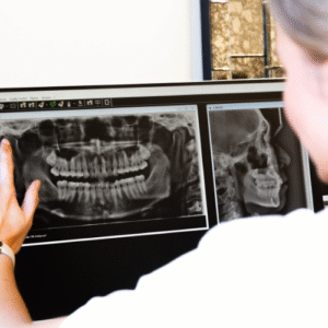
An OPG or standard CT shows the bony anatomy of the joint, but not disc position, muscle activity, airway dynamics, or the functional relationships that drive symptoms. On the radiation question, see how safe are dental x-rays and how safe are dental X-rays and when do they become unsafe?
You can look at a normal-appearing joint on X-ray in a patient whose muscles are in constant spasm, and the X-ray will tell you nothing useful.
These methods are not useless — they are insufficient for complex presentations. Objective data does not replace clinical judgement; it gives it information human senses cannot detect. The technology page lists the equipment in use here.
EMG muscle mapping: seeing what muscles are doing
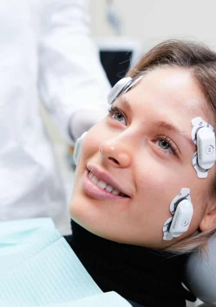
Electromyography (EMG) measures the electrical activity muscles generate when they contract. The TMD Clinic uses surface EMG — non-invasive electrodes on the skin over the muscles of mastication — to record and analyse muscle function in real time.
What is measured
Resting muscle activity. In a healthy jaw, these muscles should be relatively quiet at rest. In many TMD patients one or more show elevated resting activity, indicating chronic tension or guarding. EMG quantifies exactly which muscles are overactive, and by how much. Seven ways stress can affect your mouth covers one of the common drivers of that tension.
Functional muscle activity. During clenching, chewing and movement, muscles should fire in coordinated, symmetrical patterns. EMG shows whether the left and right masseters are balanced, whether temporalis and masseter activate in the right sequence, and whether any muscle is working disproportionately hard to compensate for dysfunction elsewhere.
Muscle symmetry. Left-right asymmetry is a hallmark of many TMD conditions. Someone clenching predominantly on the right shows significantly higher right masseter readings — which may correlate with a bite discrepancy, joint problem, or postural deviation. Where the bite itself is the discrepancy, what is malocclusion of the teeth? and treatment of malocclusion are the relevant reading.
Fatigue patterns. Recording activity over sustained clenching shows how quickly muscles fatigue and whether the pattern differs between sides, which may indicate chronic overload or compensatory loading.
How this changes treatment
Splint design moves from an educated guess to a targeted intervention. If the right masseter is hyperactive at rest while the left temporalis is compensating, the splint can be designed and adjusted for those specific imbalances. Without EMG, splint design rests on anatomical assumptions rather than functional data. How can a night guard be used to treat TMD? explains what a splint does and does not do. A splint is not the same appliance as a sports mouthguard, and the two are not interchangeable in either direction — what kind of mouth guard should I use? covers the sports version, and getting a new mouthguard — a trip to the chemist or the dentist? covers why an over-the-counter version is a different object again.
EMG also gives an objective baseline for measuring progress. After six weeks in a splint, the assessment can be repeated to quantify whether muscle activity has normalised — fundamentally different from asking whether it feels better, which matters but is subjective. Can TMD be fixed? and what is the best way to treat TMJ? set realistic expectations for that timeline.
T-Scan bite force analysis: the dynamic occlusal map
T-Scan records the location, timing and magnitude of tooth contacts during biting and chewing. A paper-thin sensor sits between the teeth, and as you close into full bite the system generates a real-time, three-dimensional force map.
What it reveals that paper cannot
Force magnitude at each contact. Not just where teeth touch, but how much force each contact carries. A contact that looks small on articulating paper might be bearing 40% of the total bite force. Why does a cracked tooth hurt so much? and how will my cracked tooth be treated? cover what happens when a tooth loses that argument.
Timing sequence. Ideally all teeth contact simultaneously, or within a few milliseconds. T-Scan reveals the sequence with millisecond precision.
Centre of force trajectory. As the teeth meet, the centre of force should move smoothly to a central, balanced position. T-Scan graphs that trajectory, exposing asymmetries and instabilities not detectable clinically.
Disclusion time. During sideways movement, the non-working side teeth should separate quickly. T-Scan measures the time precisely.
How this changes treatment — and one caveat, stated plainly
Occlusal adjustment becomes precise. Rather than grinding down high spots empirically, only the specific contacts carrying excessive force or creating timing imbalance are adjusted — minimal, targeted tooth modification. Given that enamel removed does not return, that precision matters. Why do I bite my cheek after a filling? is the everyday version of a bite that is a fraction too high.
Measuring the bite precisely is not the same as establishing that the bite causes TMD, and the weight of independent evidence is against that idea. The United States National Academies of Sciences, Engineering, and Medicine concluded in its 2020 consensus report that "occlusion should not be considered a contributing cause for the common TMDs" and that "treatment of the occlusion for TMDs also has no supporting evidence". The BMJ's 2023 clinical practice guideline for chronic TMD pain makes a strong recommendation against irreversible oral splints and a conditional one against reversible occlusal splints. Where irreversible occlusal work is proposed for jaw pain, that disagreement is worth raising before any enamel is removed — second opinions and corrective dentistry exists for that conversation.
For splints, T-Scan verifies the appliance provides even, simultaneous contacts and eliminates premature contacts or force concentrations.
CBCT imaging: the three-dimensional view
Cone beam CT gives three-dimensional imaging at a fraction of the radiation dose of conventional medical CT.
TMJ bony anatomy — detailed assessment of condylar morphology, articular eminence shape, joint space dimensions, and degenerative changes (osteophytes, flattening, erosion, sclerosis). This is what distinguishes muscular TMD, where joints may look normal, from arthrogenic TMD, where the joint itself is the problem — and it is precisely the distinction the DC/TMD paper says cannot be made reliably by clinical examination alone. Is TMD serious? and what is the difference between TMD, TMJ and bruxism? explain why that distinction changes the plan.
Condylar position — measuring joint space on both sides shows whether the condyles are seated symmetrically, or one is displaced. Where the skeletal relationship itself is the problem, what is orthognathic surgery?, jaw surgery and oral and maxillofacial surgeons are the next step.
Airway assessment. CBCT airway imaging allows the team to measure the minimum cross-sectional area of the upper airway, identify the level and nature of any narrowing, assess tongue position, evaluate soft palate length and thickness, and identify anatomical risk factors for obstructive sleep apnoea.
A correction worth making here. The link between sleep-disordered breathing and bruxism is widely asserted and is not established. A 2024 meta-analysis by Błaszczyk and colleagues in Sleep Medicine Reviews found that "the odds of SB presence in OSA did not differ from the control group (OR: 1.23, 95% CI: 0.47–3.20)", with no gradient across mild, moderate or severe apnoea. Airway imaging earns its place because obstructive sleep apnoea is serious in its own right and frequently undiagnosed — not because grinding is evidence of it. Where the airway is significantly narrow, a mandibular advancement splint may be considered instead of a standard stabilisation splint, and those two appliances do opposite things. Snoring and sleep apnoea is the service page, mouth breathing: the silent habit that's changing your face and your health covers the daytime half, and orofacial myofunctional therapy is one of the treatments that follows.
Dental and skeletal assessment — imaging of teeth, roots and surrounding bone, covering periodontal status and root pathology. See periodontal (gum) disease and periodontists.
Thermal imaging: detecting inflammation
Infrared thermal imaging captures temperature distribution across the face, highlighting increased blood flow and inflammation. It can identify acute inflammation around the TMJ, detect left-right asymmetries in blood flow, and monitor inflammatory changes over the course of treatment.
It is not diagnostic in isolation — it adds another objective data point alongside the rest.
Putting it together
The power is not in any single technology but in integrating the data streams. Complex dental cases: what happens when multiple specialists need to collaborate describes how that integration is organised, and complex dentistry is the service page.
A worked example
A patient presents with right-sided jaw pain and morning headaches. Traditional examination finds tenderness in the right masseter and clicking in the right TMJ. A standard night guard would be the obvious prescription.
The comprehensive assessment finds:
- EMG: right masseter resting activity three times higher than left; right temporalis showing compensatory hyperactivity
- T-Scan: a premature contact on the upper right second molar carrying 35% of total bite force, contacting 40 milliseconds before any other tooth
- CBCT: the right condyle shows early degenerative changes with reduced joint space; the airway is narrowed at the level of the tongue base
- Thermal imaging: elevated temperature over the right TMJ, consistent with active inflammation
That is a more detailed picture than the examination findings alone. The degenerative joint change and the airway narrowing are both findings a clinical examination could not have produced, and both change what should happen next. Night time tooth grinding and clenching and how can I stop grinding my teeth when I sleep? cover what patients notice of this at home.
The plan that follows
- A splint designed to reduce right-sided loading, based on the EMG and T-Scan data — see TMD and teeth grinding
- Referral for a sleep study, based on the airway findings
- Anti-inflammatory management, based on the thermal and CBCT joint findings. On the medication question specifically, muscle relaxants for jaw clenching: when they help, when they don't is the honest account
- Osteopathic treatment with the in-house osteopath for muscular management
- Conservative measures first, with any occlusal adjustment discussed against the evidence set out above rather than assumed
What is bruxism and how is it managed? covers the broader management picture.
Measuring whether it worked
The same technologies monitor progress objectively — EMG activity, force distribution and airway imaging can all be repeated to check for measurable physiological change rather than relying on reported improvement alone, important though that is.
When you know which muscles are overactive, which contacts are heavily loaded, whether the joint is degenerating, and whether the airway is compromised, you can treat with more information rather than less. Understanding your treatment sets out how findings and costs are presented before you commit, and second opinions and corrective dentistry is the route if previous treatment has not held.
Common questions
If the EMG shows I grind, does that mean I have a disorder?
No — and the international consensus on this changed in a way most dental websites have not caught up with.
The 2025 international consensus report on bruxism definitions, published in the Journal of Oral Rehabilitation after a meeting convened under INfORM, states the position plainly: "in otherwise healthy individuals, bruxism should not be considered as a disorder, but rather as a behaviour that can be a risk (and/or protective) factor for certain clinical consequences". It goes further: "we assess bruxism to determine its presence in conjunction with its possible consequences, rather than to diagnose it as a disorder", and "since bruxism is not a disorder, we do not diagnose it as such."
Read the word protective again, because it is not a typo. The same report notes that jaw-muscle activity "may contribute to maintaining the patency of the upper airway, thus preventing collapse" in people with obstructive sleep apnoea, that it may aid salivation in reflux, and that it may even have a positive effect on cognitive function. The consensus therefore states that "management of consequences is only needed and possible when any potential positive effect of bruxism is not compromised by the proposed management."
What follows practically is the question to ask about any proposed treatment: what consequence are we treating? Worn teeth, muscle pain, headaches on waking, a cracked restoration or a failing implant are consequences. A number on a report, by itself, is not one. If nothing is being damaged and nothing hurts, a measurement is information rather than an indication.
Is there a number that proves I am a grinder?
Not one you should accept at face value — and the field has deliberately moved away from cut-offs.
Historically the reference point was Lavigne and colleagues' 1996 sleep-laboratory study in the Journal of Dental Research, which compared 18 bruxers with 18 asymptomatic subjects and proposed research cut-offs of more than 4 bruxism episodes per hour, more than 6 bursts per episode and/or 25 bursts per hour of sleep, and at least 2 episodes with grinding sounds. Combined under logistic regression, those correctly predicted the clinical diagnosis in 81.3% of controls and 83.3% of bruxers — and the authors themselves closed by saying the criteria "need now to be challenged in a larger population, over time, and in subjects presenting various levels of severity".
They have been. The 2018 and 2025 consensus reports both state that "standard cut-off points for establishing the presence or absence of bruxism should not be used in otherwise healthy individuals; rather, bruxism-related masticatory muscle activities should be assessed in the behaviour's continuum." The 2025 report also retires the old possible/probable/definite grading, replacing it with the neutral descriptions subject-based, clinically based and device-based — because self-report, clinical examination and instruments "could conceivably assess different aspects of bruxism" rather than sitting in a hierarchy.
So if you are shown a figure and told it means you are a grinder, the fair questions are: measured how, compared with what, and what would change if the number were different. A threshold presented as a diagnosis is doing more work than the evidence supports.
Is it true that people bite far harder asleep than they can awake?
This is one of the most repeated claims in dentistry, and the study it traces back to does not say it.
Nishigawa, Bando and Nakano measured night-time bite force directly, publishing in the Journal of Oral Rehabilitation in 2001. Ten subjects wore instrumented acrylic appliances for three nights each at home, and 499 bruxism events were analysed from the 30 recordings. The results:
- Mean amplitude of a bruxism event: 22.5 kgf (standard deviation 13.0)
- Mean duration: 7.1 seconds (standard deviation 5.3)
- Highest amplitude in an individual: 42.3 kgf, across a range of 15.6 to 81.2
- Maximum voluntary daytime bite force: 79.0 kgf, range 51.8 to 99.7
- Mean ratio of night-time to daytime maximum: 53.1%, range 17.3% to 111.6%
The average night-time event reached about half of what the same person could generate awake. Only the very top of the range crossed 100%, which is what the authors meant when they wrote that night-time force "can exceed" the daytime maximum — can, in some people, not routinely does.
Three cautions the paper's own framing supports. This was ten people over three nights, measured through appliances at the first molars, so it is a mechanistic study rather than a population estimate. It measured force only and reported no clinical outcome, so it does not establish that higher force causes fractures, TMD or failed restorations. And any absolute figure quoted in newtons or PSI as a typical grinding force is going beyond this evidence.
The duration figure is arguably the more useful one: seven seconds is the average event. What loads a tooth over years is repetition, not a single heroic clench.
My symptoms come and go. Can one measurement really capture that?
This is the right question to ask, and the consensus report addresses it directly.
"Device-based sleep bruxism fluctuates considerably over time, whereas self-reported bruxism (sleep, awake) seems to be a fairly persistent trait over longer periods of time." The report notes that self-reported sleep bruxism has been shown to be a fairly persistent trait over a 20-year period in same-sex twins, and that self-reported awake bruxism recorded by smartphone stayed quite constant over six months of monitoring. Instrument readings, in other words, are the less stable of the two measures, not the more stable.
The same report is careful about what a clinical examination measures: "clinical examination does not measure bruxism itself but rather clinical signs of the motor behaviour", some of which are historical — tooth wear tells you what has happened over years, not what is happening now.
Three practical consequences. Keep your own account of the symptoms; it is data, not anecdote, and the consensus treats self-report as a legitimate assessment method rather than a poor substitute for one. Expect a single night or a single session to be a snapshot, and ask whether repeating it later would change anything. And when a baseline is repeated after treatment, ask how much of any change could simply be the normal fluctuation described above.
I do not grind my teeth together — I just hold my jaw tight. Does that count?
Yes, it has a name, and the consensus says it may matter as much as visible grinding.
The 2025 report defines four distinct behaviours. Clenching is "the act of sustained (static) tooth contact". Grinding is "dynamic tooth contacts", and — a point worth knowing — "grinding may or may not be accompanied by sounds", so a silent sleeper is not necessarily a non-grinder. Bracing is "the equivalent of clenching, but without tooth contact", and thrusting is the equivalent of grinding without tooth contact.
Those last two are the ones patients describe and clinicians historically overlooked, because they leave no wear facets and make no noise. The report's assessment: "emerging research suggests that bracing and thrusting may contribute to, among others, increased muscle fatigue and pain", and that although little research exists, they appear to be frequent behaviours.
That is precisely the situation surface EMG is suited to, because it records muscle activity rather than tooth contact. If your teeth show no wear but your jaw aches, that is not a contradiction — it is a description of bracing, and it should be assessed rather than dismissed. See TMD and teeth grinding.
Related reading
- What are the most common symptoms of TMD?
- Can TMD be fixed?
- What causes TMJ pain and how is it treated?
- What is the cause of my jaw pain?
- Why would I need to see a dental specialist?
- Specialist care
Practical details
Written by Dr Kia Pajouhesh, founding principal of Smile Solutions.
If you have been struggling with jaw pain, headaches, clicking or grinding, and previous treatment has not given lasting relief, the diagnosis may be the missing piece. The TMD Clinic offers the assessment described here — contact us to ask what it involves and what it costs. The price guide is the published starting point, and the clinical team is listed by name.
Smile Solutions, Level 1, 220 Collins Street, Manchester Unity Building, Melbourne VIC 3000 — getting here. Phone 13 13 96, or theteam@smilesolutions.com.au. Monday–Friday 8.00am–6.00pm, Saturday 8.30am–1.30pm, Sunday by appointment.
Published 16 July 2026. Individual results vary; diagnostic technology informs clinical judgement rather than replacing it. General information only; it does not replace advice from your treating practitioner.
Smile Solutions trades under ABN 28 193 514 103.
Images on This Page
-
https://www.facebook.com/tr?id=800557773428361&ev=PageView&noscript=1
(no alt text)
-
https://www.smilesolutions.com.au/wp-content/uploads/2025/10/calendar_month.svg
(no alt text)
-
https://www.smilesolutions.com.au/wp-content/themes/arcadian-wordpress-theme/assets/images/smile_solutions_logo_white.png
Smile Solutions Logo
-
https://www.smilesolutions.com.au/wp-content/themes/arcadian-wordpress-theme/assets/images/smile_solutions_logo_purple.png
Smile Solutions Logo
-
https://www.smilesolutions.com.au/wp-content/uploads/2025/11/Kia-150x150.jpg
Dr Kia Pajouhesh
-
https://www.smilesolutions.com.au/wp-content/uploads/2024/07/tfrtechnology_myowise_bite_improvement_technology_image-1-719x1024.jpeg
Myowise
-
https://www.smilesolutions.com.au/wp-content/uploads/2025/09/Wisdom-Teeth-Smile-Solutions-300x300.png
Smile Solutions Dentist Melbourne
-
https://www.smilesolutions.com.au/wp-content/uploads/2025/11/TMD-Teeth-Grinding-Smile-Solutions-300x270.png
Woman holding her face, looking concerned.
-
https://www.smilesolutions.com.au/wp-content/uploads/2026/07/Photography-and-Facial-Analysis-300x270.jpg
Photography and Facial Analysis
-
https://www.smilesolutions.com.au/wp-content/themes/arcadian-wordpress-theme/assets/images/family_owned_bussiness.png
Family Owned Business
-
https://www.smilesolutions.com.au/wp-content/themes/arcadian-wordpress-theme/assets/icons/icon--phone.svg
Smile Solutions Contact
-
https://www.facebook.com/tr? id=800557773428361&ev=PageView&noscript=1
(no alt text)
-
https://bat.bing.com/action/0?ti=25148060&tm=gtm002&Ver=2&mid=36d956cc-c635-4791-b129-0b3cd3d17387&bo=1&sid=1afd7e00ab2711f1a87cb197780dd627&vid=1afdaec0ab2711f1bdd3adbde4c0013a&vids=1&msclkid=N&pi=918639831&lg=en-US&sw=800&sh=600&sc=24&nwd=1&tl=EMG%20Muscle%20Mapping%20and%20Bite%20Force%20Analysis%3A%20How%20We%20Diagnose%20Jaw%20Problems%20-%20Smile%20Solutions&p=https%3A%2F%2Fwww.smilesolutions.com.au%2Fdental-articles%2Farticle%2Femg-muscle-mapping-and-bite-force-analysis-how-we-diagnose-jaw-problems%2F&r=&evt=pageLoad&sv=2&cdb=AQAQ&rn=559078
(no alt text)